Core Name: Neuroanatomy Electron Microscopy Core
Director: Teresa A. Milner, Ph.D.
Summary
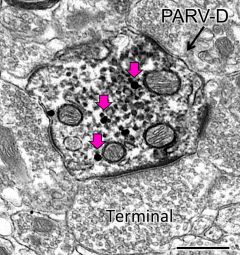
Electron micrograph from rat hippocampus shows a parvalbumin-labeled dendrite (PARV-D; peroxidase) that contains mu opioid receptor labeling (black dots; arrows) in the cytoplasm. Bar, 500 nm (Milner et al., Synapse 67: 757-772, 2013)
The goal of the Core is to provide training and services in the processing of brain tissue for light and electron microscopic immunocytochemistry. Key aspects of the core include: 1) one-on-one training classes; 2) assistance with brain fixation and histology; 3) assistance with quantitative light microscopic immunocytochemistry on brain tissue; 4) assistance with quantitative single and dual labeling electron microscopic immunocytochemistry; 5) assistance with collection, analysis and interpretation of these preparations; and 6) assistance with preparing collected data for publication and meeting presentations.
Mission of the core
This Core primarily oversees experiments on brain tissue that utilize pre-embedding light and electron microscopic immunocytochemical methods.
Location
Feil Family Research Building, 407 East 61st St, 3rd floor
Service Performed by Core
- Providing consultation with experimental design and approach
- Performing and training in perfusion fixation procedures specific for brain EM immunocytochemistry
- Training in the sectioning of brains on a vibrating microtome (Vibratome).
- Long-term storage of brain sections in cryoprotectant in -20oC freezer.
- Assisting with characterizing and determining specificities of antibodies.
- Assisting with determining optimal antibody labeling conditions for immunoperoxidase and immunogold-silver methods.
- Training in quantitative light microscopic immunocytochemical methods.
- Training in dual labeling EM immunocytochemical methods.
- Assisting with embedding brain tissue in plastic.
- Cutting thin-sections on the ultratome.
- Training in cutting thin-sections on the ultratomes.
- Training in the use of the electron microscope.
- Training in identifying the types of neuronal and glial processes on the electron microscope.
- Training in the use of specialized MCID image analysis software for analyzing electron micrographs.
- Assistance with interpretation of data and statistics.
- Assistance with preparation of data for publication and presentations.
- Assistance with writing manuscripts.
 Light micrograph shows angiotensin 1a receptor containing neurons in mouse hypothalamus 3V, 3rd ventricle Bar = 50 microns (Gonzalez et al., Neuroscience 226: 489-509, 2012)
Equipment Available in the Core
- Hitachi HT7800 Electron microscope
- 2 Leica Ultracut Ultramicrotomes
- Glass knife maker
- Oven for curing Epon plastic
- 2 Leica vibratomes
- Computer with MCID image analysis software
- Nikon light microscope equipped with NIH image software and digital camera